14 February 2019 | Enviroderm Services
We are delighted to announce the new Visioscan® VC 20plus to measure the topography of the skin.
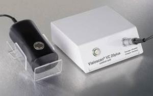
The Visioscan® VC 20plus supports a variety of claims and is an indispensable tool for the efficacy testing of cosmetics.
The Visioscan® VC 20plus is a unique UVA-light video camera with high resolution to study the skin surface directly. The SELS® parameters (Surface Evaluation of the Living Skin) have been developed especially for this camera and are used in numerous studies.The images show very impressively the structure of the skin and the level of dryness. The camera can be used on pigmented spots and lesions and on hair.
With its multi-functional software, the Visioscan® VC 20plus is a highly flexible system to characterise skin surface condition easily, accurately and very economically.
The world renowned Visioscan® in a new outfit:
- New UV-illumination source and autofocus show homogenously illuminated, high resolution, detailed skin images.
- All topography-measurements (SELS®, roughness, texture, volume and surface) are automatically calculated and displayed and saved with the image.
- New histogram-based parameters of the grey values of the image.
- Improved calculations of skin line’s directionality (anisotropy) and cell size (between the lines).
- Additionally, you can draw lines (e.g. to assess hair length or −thickness) and free-hand objects (e.g. to assess spots and lesions) regarding length, circumference, and average grey value.
- Attractive 3D-images.
- Impressively analyse Sebufix® and Corneofix®.
- Press one button to export all study data to Excel®.
- Please ask for exchange offers on the previous model Visioscan® VC 98.